![]()
Vascular Imaging/Interventional Radiology(IVR)
May 28,2024 Update
Introduction to the examination room
Vascular imaging is performed by inserting a thin tube (catheter) through an artery, such as at the base of the leg (the femoral artery), the elbow (brachial artery), or the wrist (radial artery). The catheter is advanced to the desired blood vessel by viewing the progress in a fluoroscopic image (X-ray), and a contrast agent is injected in order to see the movement and condition of the blood vessels as well as to dye tumors. This term is sometimes shortened and called angio.
In recent years, we have been performing more endovascular treatments, image-based treatment (Interventional Radiology: IVR) to inject anti-cancer drugs to treat cancer, expand the narrowed parts of blood vessels, inserting an embolic substance to block blood flow at a bleeding site, cancer or aneurysm, instead of performing surgical operations.
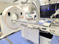
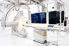
There are three vascular imaging devices operating in the vascular imaging rooms of the Department of Radiology; a vascular imaging device with a focus on the head and neck area (PHILIPS Allura Clarity FD20/15), a vascular imaging device with a focus on the cardiac area (PHILIPS Azurion7 B12), and an IVR-CT device (SIEMENS: Nexaris Angio-CT) that covers the entire body area.
The Vascular Imaging Division is engaged in examination and treatment by physicians, nurses, clinical radiologists, and clinical engineering technicians (for the cardiac area), working together as a team.

Time for examination/Important points
Most examinations and treatments are performed in the hospital. Pre-treatment and time will vary depending on the nature of the examination/treatment.
The details will be explained upon request by the physician or nurse. If you have any questions, please ask them.
Contact information
049‐228‐3519 [Department of Radiology, Vascular Imaging Room Direct]