![]()
General Radiography/Mammography
July 3,2024 Update
General Radiography/Mammography
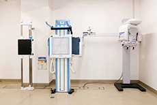
General radiography is an examination where the amount of X-ray radiation transmitted through the human body is expressed as a black and white image. This is also commonly referred to as x-ray photography or taking pictures of the bones in general, and soft tissues (such as breast imaging), including the chest, abdomen, head and neck, spine, and limbs, etc. The imaging rooms are on the first floor in Radio-Imaging Diagnostics, and imaging rooms X-1 to X-5 are used.
Introduction to the imaging rooms
Applications and features of each imaging room
| Imaging Room | Application |
|---|---|
| X-1 | Mainly used for chest and abdominal imaging. |
| X-2 | This is a wide-angle imaging room that can be used for multiple purposes. It is used for chest and abdominal imaging, as well as bones in general and specific imaging, etc. |
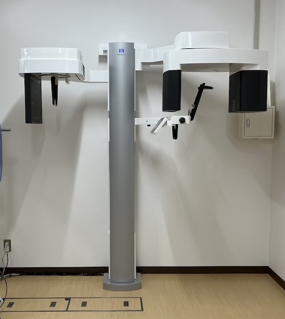
| X-3 | This is a wide-angle imaging room that can be used for multiple purposes. In addition, there is a panoramic X-ray unit, and images for the oral surgery area can be obtained. |
| X-4 | This is a wide-angle imaging room that can be used for multiple purposes. It is used for chest and abdominal imaging, as well as bones in general and specific imaging, etc. |
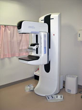
| X-5 | Mainly used for breast imaging (mammography). |
The systems in each imaging room
| Imaging Room | Equipment |
|---|---|
| X-1 | Canon Medical Systems X-Ray Generator KXO-50SS Konica Minolta AeroDR System (fine) Konica Minolta Diagnostic Imaging workstation CS-7 Nonaka Manufacturing Co., Ltd. Wall-mounted reader imaging table 11BZ0546 Obayashi Manufacturing Co., Ltd. Reader imaging stand ROCKET EVOLUTION Obayashi Manufacturing Co., Ltd. Lifting-type floating imaging table E・JIS |
| X-2 | Canon Medical Systems X-Ray Generator KXO-50SS (2 Tubes) Konica Minolta AeroDR System (fine) Konica Minolta Diagnostic Imaging workstation CS-7 Sankyo Medical Co., Ltd. Long reader imaging table SA-R1 Obayashi Manufacturing Co., Ltd. Reader imaging table ROCKET EVOLUTION Obayashi Manufacturing Co., Ltd. Reader imaging table ROCKET EVOLUTION (for long strokes) Obayashi Manufacturing Co., Ltd. Lifting-type floating imaging table E・JIS |
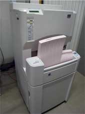
| X-3 | Canon Medical Systems X-Ray Generator KXO-50SS (2 Tubes) Konica Minolta AeroDR System (fine) Konica Minolta Diagnostic Imaging workstation CS-7 Konica Minolta Image reading system (CR) REGIUS MODEL 190 Sankyo Medical Co., Ltd. Long reader imaging table SA-R1 Obayashi Manufacturing Co., Ltd. Reader imaging table ROCKET EVOLUTION Obayashi Manufacturing Co., Ltd. Vertical floating imaging table E・JIS MORITA panoramic x-ray scanner Veraview X800 |
| X-4 | Canon Medical Systems X-Ray Generator KXO-50SS (2 Tubes) Konica Minolta AeroDR System (fine) Konica Minolta Diagnostic Imaging Workstation CS-7 Sankyo Medical Co., Ltd. Long reader imaging table SA-R1 Obayashi Manufacturing Co., Ltd. Reader imaging table ROCKET EVOLUTION Obayashi Manufacturing Co., Ltd. Vertical floating shooting table E・JIS |
| X-5 | Fujifilm Healthcare Systems X-Ray Generator Radnext50 HOLOGIC Breast Imaging Equipment Selenia Dimensions (tomosynthesis-compliant) HOLOGIC Diagnostic Support System Cenova Konica Minolta AeroDR system Konica Minolta Diagnostic Imaging Workstation CS-7 Obayashi Manufacturing Co., Ltd. Vertical floating imaging table New SUD-3 |
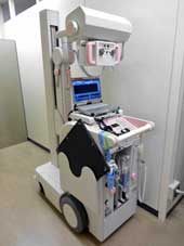
| Mobile X-Ray Equipment |
Fujifilm Healthcare Systems Mobile X-Ray Equipment Sirius 130HP, Sirius Starmobile tiara airy Konica Minolta AeroDR Konica Minolta Portable Solution System (CS-7 Portable) |
| Image inspection | Konica Minolta I-PACS QA (with IP-pro) |
At our hospital, mammography is performed by clinical radiologists who are certified in screening mammography for medical checkups in order to provide more accurate imaging. Our equipment is a device that is equipped with a breast tomosynthetic system.
With a conventional two-dimensional (2D) imaging, a lesion may be hidden in the mammary gland tissue, or it may not be diagnosed due to the overlap of mammary gland tissue.
Tomosynthesis (3D) is a three-dimensional imaging method that can reduce the overlap of the mammary gland and allow visualization of lesions more clearly.
- Those who are pregnant or may be pregnant
- Those who have breast implants
- Those who have a pacemaker implanted
- Those who are breastfeeding (because the pressure can easily make the milk flow)

Important Points
- Please talk to your doctor or the technician in charge if you are pregnant or have a possibility of being pregnant.
- There are basically no issues for areas outside the area to be imaged, but there are some items that may affect if worn during imaging. The following objects are to be removed, and you may have to change into examination clothes.
- Metal products (necklaces, rings, watches, earrings, dentures, magnetic patches, bras, etc.)
- Buttons, poultices, hand warmers, T-shirts (with rubber prints, lamé (interwoven metal), wet diapers, etc.)
- Corsets, supporters, etc.
- In addition to the above, please feel free to contact us if you have any questions.
Q & A for general radiography
- Q How long will the imaging take?
- The imaging time depends on the part of the body being imaged and the method of imaging.
Also, please understand that the order of events may differ depending on the patient's condition. - Q Why do you shoot so many images at a time?
- It is common to evaluate the angle and the direction in the body by obtaining multiple images with different views in order to make an accurate diagnosis.
- Q Is it okay to take multiple images in a short period of time?
- In the case of the patient whose progress is being observed, there may be a case where we need to take a picture every time there is an outpatient examination, and there may be cases where we would take images daily, depending on the symptoms.
The amount of radiation used for the imaging is very small, but if you feel uneasy, please consult directly with your doctor or the attending physician. - Q I hear mammography is painful, but is it really okay?
- The pressure plate is set so that it does not press with more than a set pressure. Please feel free to talk with us if you have any questions. Please tell your technician if you feel a strong pain or anxiety.
- Q What are some things to be mindful of when getting a mammogram?
- In mammography imaging, we will image the parts including from the armpit to the breast. Because antiperspirant, powder, and lamé can be reflected in the image, please wipe these well before taking the image. Antiperspirants and powders can appear very similar to small calcifications and masses. Please refrain from applying antiperspirant or powder on a day when you will be having an image taken.