![]()
X‐ray Fluoroscopy
June 5,2024 Update
X‐ray Fluoroscopy
Some structures in the human body are visible with x-rays, while other structures are not. With fluoroscopy, a contrast agent is injected into the gastrointestinal tract or veins, etc., and the morphology of structures that are not normally visible with x-rays can be inspected. This is called a contrast examination.
The examination rooms are in Radio-Imaging Diagnostics on the first floor, and the examination is done in examination rooms from X-8 to X-10.
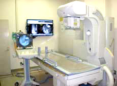
Introduction to the X‐ray fluoroscopy rooms
Equipment and applications of each examination room
| Examination room | Equipment | Application |
|---|---|---|
| X-8 | Canon Medical Systems ZEXIRA DREX-ZX80/P6 | Obstetrics and gynecology, urology, orthopedics surgery, pediatrics, etc. |
| X-9 | Canon Medical Systems ZEXIRA DREX-ZX80/06 | Obstetrics and gynecology, urology, orthopedic surgery, pediatrics, etc. |
| X-10 | Shimadzu Corporation SONIALVISION G4 | Upper gastrointestinal tract contrast examination, lower gastrointestinal tract contrast examination, obstetrics and gynecology, urology, orthopedic surgery, pediatrics, etc. |
| X-6 |
Dornier MedTech Extracorporeal shock wave lithotripter DELTA III PRO |
Urinary tract calculus/ biliary tract calculus lithotripsy |


Typical examination in an X-ray Fluoroscopy exam room
Upper gastrointestinal tract examination (esophagus/stomach)
This is an examination to observe the condition of the esophagus, the shape of the stomach and mucous membranes that is conducted by drinking barium (contrast agent).
Target symptoms: Neck discomfort, heartburn, epigastric pain, stomach discomfort, nausea, vomiting, etc.
Target diseases : Esophageal cancer, reflux esophagitis, gastric cancer, gastric ulcers, etc.
[Time for the examination]
The examination time is about 20 minutes.
Lower gastrointestinal tract examination (colon)
This is an examination where they inject barium and air via the anus and observe the shape of the colon and the condition of the mucous membranes.
Target symptoms: Diarrhea, constipation, abdominal bloating, bloody bowel discharge, stool occult blood positive, etc.
Target diseases : Colon cancer, colon polyps, ulcerative colitis, Crohn's disease, etc.
[Time for the examination]
The examination time is about 30 minutes.
Urological examination: Intravenous urography (IVU)
This is an examination to observe the shape and function of the kidneys, ureter, and bladder by injecting an iodine contrast agent into a vein in the arm and taking an X-ray of the abdomen.
Target symptoms: Hematuria, back pain, etc.
Target diseases : Kidney stones, ureter stones, kidney cancer, urinary tract cancer, bladder cancer, hydronephrosis cause search, etc.
[Time for the examination]
The examination time is about 30 minutes.
Hysterosalpingography (HSG)
This is mainly an examination done in the search for causes of infertility. An iodine contrast agent is injected into the endometrial cavity to observe the shape of the endometrial cavity and the passage condition of the fallopian tubes.
[Time for the examination]
The examination time is about 20 minutes.
Extracorporeal shockwave lithotripsy (ESWL)
This is a treatment method to crush stones from outside the body without surgical treatment of the stones (mainly urinary tract stones) and to eliminate the stones from outside the body.
X-rays or ultrasound are used to aim at the stones.
You may feel a dull pain during the treatment.
[Treatment Time]
The treatment time is about 60 minutes.
Important Points
- Please talk to your doctor or staff if you are pregnant or have a possibility of being pregnant.
- Be sure to remove bras, buttons, magnetic patches, etc. and change into exam clothes.
- Please contact the outpatient clinic if you have any questions regarding changes to your appointment.
- Please feel free to contact us if you have any questions about the examination.
Q&A about X-Ray Fluoroscopy
- Q. How much barium do you drink for imaging the upper gastrointestinal tract?
- The amount of barium is about 100 – 150 cc, which is less than in the past, but it depends on the size and shape of the stomach.
- Q. Why do I eat the examination meals for the lower gastrointestinal tract examination?
- We may not be able to inspect the large intestine, or you may not get the correct test results if there is stool left in the colon, so you need to eat so that as little stool will remain as possible. This will affect the accuracy of the exam.
- Q. What happens to the contrast agent that is used?
- When using barium, it will be excreted in the feces.
- When an iodine contrast agent is used, it is excreted in the urine.
- Patients with no fluid restrictions should try to consume fluids after the exam.
- When an iodine contrast agent is used, it is excreted in the urine.